Morris Eye Group
  • About Us
    • Our Doctors >
      • Jeffrey B. Morris, M.D.
      • Ardalan Eddie Aminlari, M.D.
      • Eric W. Johnston, M.D.
      • Christina Kytasty, D.O.
      • Connie Tan, O.D.
      • Mona Moshtaghi, O.D.
    • Our Practice & Facilities
    • Patient Testimonials
    • Our Technology
    • In The News
    • Join Our Team
    • Blog
    • CDC COVID Symptom Test
  • Services
    • Cataract Center
    • Dry Eye Center
    • LASIK Center
    • Glaucoma Center
    • Cornea Center
    • Optical Center
    • Aesthetic Center
    • Subspecialist
    • Comprehensive Eye Exam >
      • Refractive Errors
      • Pediatric Eye Exams
      • Diabetic Eye Disease
      • Macular Degeneration
      • Retinal Detachment
      • Floaters & Flashes
      • 10 Tips For Healthy Eyes
  • Patient Information
    • Schedule Appointment
    • Patient Education
    • Patient Forms
    • Patient Portal
    • Pay a Bill
    • Insurance
    • CareCredit Financing
    • Flex Spending & Tax Resources
    • Surgery Center Information
    • Cataract Pre and Post Surgery Videos
  • Online Store
    • Contact Lenses
    • Macuhealth
    • Macuhealth Plus
  • Contact Us

Oh no, I think I have Pink Eye!

3/28/2022

6 Comments

 
You may have pink eye or another problem with your eyes.  Here are the symptoms of Pink Eye: pink or red eyes, itchy, burning, watery eyes, a discharge coming from the eye (either white, yellow or green) and crusting along your eyelids that can even prevent your eyes from opening.
Pink Eye is also known as conjunctivitis and that can either be viral or bacterial.

 A viral infection is likely to start in one eye and spread to the other. If you are having a clear, watery discharge from your eyes and cold-like symptoms, you are likely having a viral infection.  This infection will likely last 7-10 days.

If it is a bacterial infection there may also be cold-like symptoms however, the notable difference is the thick (puss-like) discharge that forces your eyes to stick together.  Bacterial pink eye lasts roughly 3-5 days and requires antibiotic eye drops to treat the infection.

Pink Eye does spread very easily.  The main action to prevent spreading pink eye is to wash your hands, whether you are the patient or you have a family member or friend with pink eye.  If infected, avoid touching or rubbing your eyes.  Avoid sharing personal items with other such as linen, pillow cases, towels, make-up or glasses.
​
Most cases of viral pink eye do go away on their own, but you may need to be treated if you are in a lot of pain, noticing blurry vision or if you are sensitive to light.  Bacterial pink eye should be treated by your eye doctor.  If you are a contact lens wearer and you have pink eye, stop wearing your contact lenses till your treatment is complete or the symptoms have completely disappeared.  Are you a contact lens wearer?   You should always have a pair of back-up glasses with a current prescription for situations such as pink eye.
6 Comments

FDA approves a contact lens for treatment of myopia in children

7/30/2021

9 Comments

 
Picture
There is a new treatment for eyecare professionals to manage myopia progression. Made by Coopervision, MiSight is a daily disposable soft contact lens. It is the only method approved by the FDA to slow the progression of myopia in children (8-12 years of age at the initiation of treatment).

Our very own Dr. Tan is one of the few optometrist certified to fit this lens that helps control myopia in children. 


Myopia, or nearsightedness, is an eye condition in which the eyeball elongates during development, thus causing blurry vision since the light rays focus in front of the retina (instead of correctly onto the retina). In 2010, 28% of the world’s population was myopic - by 2050, that number could reach 50%. With the growing myopia epidemic, there has been a greater need to slow down the progression of myopia. With higher myopia, there is a greater risk of developing other eye diseases such as glaucoma, retinal detachment, cataracts, and myopic maculopathy. Furthermore, the patient will also be more dependent on glasses and it may be more difficult to be a good candidate for refractive surgery. 

The increasing incidence of myopia can be attributed to a combination of two factors: genetics and environment.

For genetics, even one parent with myopia increases the child’s chance of developing myopia by 3x, which can double to 6x if both parents are myopic.

For the environment, research has shown that spending more time
outdoors lowers the risk of progressive myopia.

The earlier myopia management begins, the better the outcomes will be regarding the child’s overall eye health. Managing myopia starts with annual comprehensive eye exams. 

In the 3 year randomized clinical trial, the Misight lens decreased a child's average prescription from progressing by 59% and preserved a child's eye length by 52% versus the control lens. There were no adverse effects with contact lens wear and the kids found it comfortable to wear.

Not only does Misight treat myopia progression, it corrects the child’s prescription and allows them to be independent of glasses (which is great for active patients). 


9 Comments

Dry Eyes?

1/14/2020

565 Comments

 
Picture
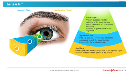
What is Dry Eye Disease?
Dry Eye Disease or Meibomian Gland Dysfunction is much more commonly seen than in previous decades.  Those that have it know its symptoms well: dryness, watery eyes, itchiness, stinging, burning, grittiness, redness, blurry vision and a foreign body sensation in the eye.  The symptoms just don’t seem to go away and are very annoying.  More than annoying, dry eyes can also produce chronic eye inflammation, corneal abrasions and corneal ulcers. Patients sometimes come in complaining that they have blurry vision and think they need new glasses when in fact, they have Dry Eye Disease that is disrupting their vision.

Is it a simple problem to fix?
Yes and No.  Firstly, it requires an eye doctor to take a careful history, medical review of existing diseases and an eye examination. Correct Diagnosis of the cause is very important as it will dictate what treatments will work and what will not. If a patient has contact lenses that are not fit well, it may be a simple fix to change the contact lens prescription.

How is Dry Eye Disease treated?
Early treatment is the key to preventing irreversible damage to the glands of the eyes. The doctors of Morris Eye Group have a three- prong treatment plan with the goal to not just treat the symptoms but to treat the underlying cause.  This necessitates a proper diagnosis of the underlying problem.

Our doctors analyze the patient’s tear film quantity and quality.  Are the eye glands producing proper lubrication to the eye or is the lubrication lacking enough essential oils to do the job?   If the dry eye is a side-effect of a prescribed medication our doctors work with a patient’s primary care doctor or specialist to find an alternate medication to those that produce dry eyes.  If a patient’s Meibomian glands are blocked the doctors prescribe a treatment to unblock them.  There is also a home regimen to keep the glands open and a new Omega7 nutritional supplement that promotes the production of the oils that bathe the eye.   We always recommend adding UV protection in prescribed eye wear.

What causes Dry Eye Disease?
 The causes of Dry Eye Disease are varied and many.  Here are the main causes:
Aging, contact lens wear, autoimmune diseases such as Lupus, Sjorgen Syndrome and Thyroid Disease. Many prescribed drugs produce dry eye side-effects.  We also know that computer, tablet and smart phone use have added to eye strain from their screen- emitted blue light.  
It is more common in females and in those that participate in outdoor sports such as golf, surfing and tennis.  Dry Eye may also be due to an occupational exposure to known eye irritants.   In some instances, over -the -counter eye drops to clear up red eyes actually make Dry Eye Disease worse.  ​

565 Comments

Diabetic Eye Disease Q & A

10/29/2019

769 Comments

 
Picture

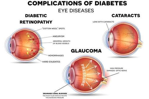
How does diabetes damage your eyes?
Diabetes can bring with it a host of health problems, and among other things, it is known to affect our eyes. High blood sugar can lead to problems like blurry vision, cataracts, glaucoma, and retinopathy. In fact, diabetes is the primary cause of blindness in American adults ages 20 to 74. The most common diabetic eye disease is Diabetic Retinopathy, and it is estimated to affect almost 11 million Americans by 2030.

​
Diabetic Retinopathy is caused by damage to the blood vessels of the light-sensitive tissue at the back of the eye (the retina), and can lead to serious vision problems including:
​
Vitreous hemorrhage: The new blood vessels may bleed into the clear, jelly-like substance that fills the center of your eye. If the amount of bleeding is small, you might see only a few dark spots (floaters). In more-severe cases, blood can fill the vitreous cavity and completely block your vision. Vitreous hemorrhage by itself usually doesn't cause permanent vision loss. The blood often clears from the eye within a few weeks or months. Unless your retina is damaged, your vision may return to its previous clarity.
Retinal detachment: The abnormal blood vessels associated with diabetic retinopathy stimulate the growth of scar tissue, which can pull the retina away from the back of the eye. This may cause spots floating in your vision, flashes of light or severe vision loss.
Cataracts: You're more likely to have cataracts, and at a younger age, if you have diabetes. The lens of the eye gets oxygen and glucose from the aqueous humor, which is the fluid that fills the front of the eye. If someone doesn't have control over their glucose levels, such as someone with diabetes, the sugar levels can rise, causing the lens to swell and consequently the early development of cataracts.
Glaucoma:  Having diabetes doubles your odds of glaucoma. New blood vessels may grow in the front part of your eye and interfere with the normal flow of fluid out of the eye, causing pressure in the eye to build up. This extra pressure can damage the retina and the optic nerve, the main nerve for sight that carries images from your eye to your brain, causing permanent vision loss.
Blindness: Eventually, diabetic retinopathy, glaucoma or both can lead to complete vision loss.


What are symptoms of diabetic eye diseases?
At first, in the early stages of diabetic eye disease, there may be no symptoms or only mild vision problems. However if gone untreated, eventually diabetic eye disease can cause blindness.
 
As the condition progresses, symptoms may include:
-Spots or dark strings floating in your vision (floaters)
-Blurred vision
-Fluctuating vision
-Impaired color vision
-Dark or empty areas in your vision
-Vision loss  

Should you get yourself screened for diabetic eye disease?
If you are 12 years or older and have been diagnosed with diabetes, it is recommended that you see your eye doctor yearly for an eye exam with dilation (even if your vision seems fine). Diabetic eye disease can develop in anyone who has type 1 or type 2 diabetes. While diabetes doesn't necessarily always lead to vision loss, taking an active role in diabetes management can go a long way toward preventing diabetic retinopathy.
The risk of developing a diabetic eye disease can increase as a result of:

-Duration of diabetes: the longer you have diabetes, the greater your risk of developing diabetic retinopathy
-Poor control of your blood sugar level
-High blood pressure
-High cholesterol
-Pregnancy
-Tobacco use
-Genetics: especially for those of African-American, Hispanic or Native American descent  
 
How are diabetic eye diseases diagnosed?
Diabetic eye disease is best diagnosed with a comprehensive dilated eye exam. For this exam, dilating drops placed in your eyes widen your pupils to allow your doctor to better view inside your eyes to check for any signs of diabetic damage.
During the exam, your eye doctor will look for:

-Abnormal blood vessels
-Swelling, blood or fatty deposits in the retina
-Growth of new blood vessels and scar tissue
-Bleeding in the clear, jelly-like substance that fills the center of the eye (vitreous)
-Retinal detachment
-Abnormalities in your optic nerve
In addition, your eye doctor may:
-Test your visual acuity
-Measure your eye pressure to test for glaucoma
-Look for evidence of cataracts
-Request an optical coherence tomography (OCT) test. This imaging test provides cross-sectional images of the retina that show the thickness of the retina, which will help determine whether fluid has leaked into retinal tissue. And later, OCT exams can be used to monitor how treatment is working.  

How do you treat diabetic eye diseases?
​Treatment, which depends largely on the type of diabetic eye disease you have and how severe it is, is geared to slowing or stopping progression of the condition. If you have mild or moderate diabetic retinopathy, you may not need treatment right away. However, your eye doctor will closely monitor your eyes to determine when you might need treatment. If you have advanced, or proliferative diabetic retinopathy or macular edema, you'll most likely need prompt surgical treatment by a retina specialist. Depending on the specific problems with your retina, options may include: laser treatments, surgical vitrectomy, injections, intravitreal steroids, and/or other medications.
 
Even after treatment you'll still need regular eye exams. And at some point, additional treatment may be recommended. Surgery often slows or stops the progression of diabetic eye disease, but it's not a cure. Because diabetes is a lifelong condition, future retinal damage and vision loss are still possible; thus it is imperative to work closely with your ophthalmologist, primary care doctor, and nutritionist to control your diabetes long-term.
 
Can I prevent diabetic eye diseases?
Unfortunately, we can't prevent diabetic eye disease which is why careful management of your diabetes and early intervention is the best way to prevent vision loss.

​If you have diabetes, reduce your risk of getting diabetic eye disease by doing the following:

 
Manage your diabetes: Make healthy eating and physical activity part of your daily routine. Try to get at least 150 minutes of moderate aerobic activity, such as walking, each week. Take oral diabetes medications or insulin as directed.
Monitor your blood sugar level: You may need to check and record your blood sugar level several times a day — more-frequent measurements may be required if you're ill or under stress. Ask your doctor how often you need to test your blood sugar.
Ask your doctor about a glycosylated hemoglobin test: The glycosylated hemoglobin test, or hemoglobin A1C test, reflects your average blood sugar level for the two- to three-month period before the test. For most people, the A1C goal is to be under 7 percent.
Keep your blood pressure and cholesterol under control: Eating healthy foods, exercising regularly and losing excess weight can help. Sometimes medication is needed, too.
If you smoke or use other types of tobacco, ask your doctor to help you quit: Smoking increases your risk of various diabetes complications, including diabetic retinopathy.
Pay attention to vision changes: Contact your eye doctor right away if you experience sudden vision changes or your vision becomes blurry, spotty or hazy. Pregnancy may worsen diabetic retinopathy, so if you're pregnant, your eye doctor may recommend additional eye exams throughout your pregnancy.  
 

References:
Ahdieh, H. (2018, October 30). Diabetes And Eye Disease. Retrieved October 22, 2019, from https://www.lvcenter4sight.com/diabetes-and-eye-disease/.
Diabetic retinopathy. (2018, May 30). Retrieved October 22, 2019, from https://www.mayoclinic.org/diseases-conditions/diabetic-retinopathy/symptoms-causes/syc-20371611.

769 Comments

Home Eye Safety

9/27/2019

15 Comments


How to protect your eyes outdoors
The sun releases energy (radiation) in many forms. The sunlight we see, the heat we feel, and ultraviolet (UV) rays. UV rays can cause sunburn and damage to your eyes. There are two types of UV rays both invisible to the eye; UV-A and UV-B rays. UV-A rays can cause harm to your central vision by damaging your macular. The macula is part of the retina at the back of your eye. UV-B rays effect the front part of your eye (the cornea and the lens). While both UV-A and UV-B rays can cause damage too your eyes UV-B does cause more. When you’re exposed to an excessive amount of UV-rays and have minimal eye protection you run the risk of developing Macular Degeneration, Cataracts, Pterygium, Skin Cancer, and Corneal Sunburn. 

The two best ways to avoid harmful UV rays is to understand the dangers and to wear proper eye protection that blocks the UV rays. While these rays radiate directly from the sun they also reflect from the ground, water, snow, sand, and other bright surfaces. This leaves everyone, including children, at risk of developing eye damage from UV radiation. If you are someone who spends long periods of time out in the sun you are at the greatest risk. 

Choosing proper eye protect can help filter light and protect your eyes from the harmful UV radiation. When deciding on a pair of sunglasses there are multiple things that you should look for. Sunglasses that reduce glare, filter out 99-100% of UV rays, protect your eyes, are comfortable, and do not distort colors. Children are no exception to UV radiation exposure and should also make sure they have protective eyewear. 
​

For certain outdoor activities, you may also have to use eyewear designed to provide impact protection for that activity. Please see the list below of a few outdoor activities and consider the type of eyewear protection you might need. 

Biking/Cycling: UV protection and protection from wind and debris
Boating: UV and glare protection
Skiing/winter sports: UV protection, polarized lenses to reduce glare and brightness, and yellow, amber, orange-red tints to improve contrast 
Hiking/Mountain Climbing: UV protection and polarized lenses to reduce glare
Racquet/Ball Sports: Protection eyewear and UV protection 
Working Outdoors with Powertools/Chemicals: Protective eyewear and UV protection. 

Protect Your Eyes from the Sun. (2019). Retrieved September 27, 2019, from https://www.preventblindness.org/protect-your-eyes-sun.
 

Proper Contact Lens Hygiene
Proper care is necessary for successful contact lens wear, normal lens life, and good eye health. Your doctor will provide you with products to clean, disinfect, and store your lenses. 
To be fit for contact lenses our office requires that our patients complete all three components of the eye examination: medical exam, refraction, and contact lens measurements. Even if one is an experienced contact lens wearer all three components of the examination must be completed. Patients are provided with their contact lenses during their initial fitting.

New Patients and New Contact Lens Wearers:
Contact lenses are medical devices that are placed on the eye. It is important that we determine that they are properly fit, that they are not compromising corneal health, and that there are no other problems related to the wearing time or infection. Once ordered, our opticians will spend time educating you on insertion, removal, proper care and your own specific wearing regimen. Once dispensed, we will be seeing you for a series of follow-up appointments over the next 90 days to confirm that you are correctly fit.
After 90 days, you may order replacement lenses from us or any other vendor of your choice. We will be happy to provide you with a contact lens prescription once we are confident you are correctly fit. Contact lens prescriptions are good for one year.

We recommend that all of our contact lens patients have a pair of glasses on hand. After your refraction you will be given a copy of your glasses prescription. This prescription is good for two years. Additionally, wearing sunglasses is an important product to one’s eyes from UV rays. Sometimes contact lenses make one more sensitive to bright light. If you only wear contact lenses occasionally, then you will need a pair of prescription sunglasses.

Established Patients and Long-Time Contact Lens Wears:
If you currently wear contact lenses, we recommend an annual medical eye examination to ensure the health of the eye and the fit of the lenses. Moreover, from year to year there is often a change in vision and there may need to be a correction made in your contact lens or glasses prescriptions. If there is a significant change in your refraction, a second contact lens examination may be required by our contact lens specialist. The process for fitting and dispensing an updated pair of contact lenses is identical to the initial process described previously. 
Once this process is complete, we would once again be pleased to have you reorder your lenses through our dispensary or provide you with your contact lens prescription to be used with any other vendor. 

BASIC DO’S AND DON’TS
DO use only solutions recommended by your doctor.
DO check for lens damage before each wear.
DO replace your contact lens case once a month to avoid infection.
DO keep all of your follow-up appointments and annual eye exams.
DO wear sunglasses with proper UV protection.
DO have a spare pair of glasses on hand.
DO carry your prescriptions when traveling.
DO call us if you have questions or problems.
DO call to reorder before you run out of disposable lenses.

DON’T wet your lenses with your saliva.
DON’T rub your eyes when wearing your lenses.
DON’T insert a lens from a packet that is damaged.
DON’T forget to clean and disinfect non-disposable lenses.

The solution give to you by your provider is prescribed specifically for your lenses and eyes. Since they can vary significantly from one manufacturer to another, do not change or substitute brands unless you check with your doctor first. Use of improper solutions may result in lens damage or eye irritation. 
The adaptability of your eyes is the key factor in determining wearing time. Do not exceed the wearing. Do not exceed the wearing schedule that is prescribed for you. 
Remember, like any medical device, contact lenses must be monitored on a regular basis. Professional follow up care is the most important element in successful long-term lens wear. Please keep your scheduled appointments. 

In the beginning it is normal if: 
1) Your eye itch or feel funny
2) One lens is more noticeable then the other
3) Your vision seems fuzzier than with glasses 
4) You have trouble handling your lenses
Remove your lenses immediately if:
1) You develop unusual pain or redness
2) You develop unusual foggy or cloudy vision
3) You experience a decrease in vision
4) You suspect something is wrong


Protecting your eyes while on the computer 
Blue light is a color in the visible light spectrum. Blue light is everywhere in our world and it a short wavelength that produces high amounts of energy. The sun use to be the only source of blue light. However, now we have brought blue light inside by the way of digital screens (found on TV’s, smartphones, computers, laptops, tablets, and gaming systems), electronic devices, LED and fluorescent lighting. 

You can minimize the amount of blue light that enters your eyes by wearing glasses the filter some of these harmful rays. Morris Eye Group offers a high-quality blue light lens that eliminates a portion of the short wavelength. 

Children’s eyes are particularly vulnerable to the effects of blue light as virtually all of the blue light they are exposed to passes through their cornea and lens and reaches their retina. Coupled with a high amount of screen time, this puts them at risk for over-exposure to blue light that may prove to have long-term consequences. Recommended below are some safe technology habits for you and the entire family: 

• Say goodnight to technology two hours before bedtime. 
• Limit children’s screen time to one to two hours per day for children over two years of age and restricting it completely for children under two years old. 
• Turn down the brightness of your device. 
• Change digital device background colors from bright white to warmer colors to reduce 


Importance of Artificial Tears
Artificial tears are considered a first-line therapy for dry eye. They increase tear volume and lubricate the ocular surface. They often provide temporary relief of irritation symptoms in many dry eye conditions.
Tears can be used on an as-needed basis by most dry eye patients. Patients with severe conditions should be instructed to use artificial tears on a regular basis (every 1–2 hours) and to increase the frequency of instillation when reading or when they are exposed to dry or drafty environments, until a more definitive treatment can be instituted.
Stephen C. Pflugfelder, Gregory R. Nettune, in Ocular Surface Disease: Cornea, Conjunctiva and Tear Film, 2013

Most common causes of eye injuries to children at home
Each year, thousands of children age 14 and younger suffer serious eye injuries, even blindness, from toys. Some of the most common eye injuries children experience at home are: 

• Misuse of toys 
• Falling from their bed or into furniture
• Misuse of everyday tools and objects 
• Contact with harmful household products 
• Automobile accidents.

Below are a few safety tips to help protect your children indoors: 
- Use safety gates at the top/bottom of stairs
- Provide lights/handrails to improve safety on stairs 
- Pad/cushion sharp corners and edges of furnishing and home fixtures
- Install cabinet and drawer locks in kitchen and bathrooms 
- Store personal-use items (cosmetics, toiletry products), kitchen utensils, and desk supplies out of reach for children
- Keep paints, pesticides, and fertilizers, and similar products properly stored in a secure area 

Below are a few safety tips to help protect your children with their toys: 
- Read all warnings and instructions on toys
- Avoid toys with sharp or rigid points, shafts, spikes, rods, and dangerous edges
- Keep toys intended for older children away from younger children 
- Avoid flying toys and projectile-firing toys; that pose potential eye hazards 
- Keep BB guns away from kids 
Protecting young children from eye injuries at home and at play. (2019). Retrieved September 27, 2019, from https://www.preventblindness.org/protecting-young-children-eye-injuries-home-and-play.

Hazardous activities at home that you should be wearing protective eye wear for
Wearing protective eyewear can help prevent 90 percent of eye injuries. It is important to make sure that your home has at least one approved pair and that you and your family members wear the eyewear when involved in hazardous activity.
There are over 125,000 eye injuries each year using common household products such as oven cleaner and bleach for cleaning and other chores. When you are cooking foods that can splatter hot grease or oil you should make sure you have protective eyewear. 
Other hazardous activities you should protect your eyes from are: 
- Drilling or hammering screws or nails into walls or hard surfaces like brick or cement
- Mowing the lawn.
- Using a power trimmer or edger.
- Using tools (power or hand).
- Working with solvents or other chemicals.
- Any task that can produce fragments, dust particles or other eye irritants.
Eye Injuries at Home. (2016, April 14). Retrieved September 27, 2019, from https://www.aao.org/eye-health/tips-prevention/injuries-in-home.

15 Comments

Healthy Eye Aging

9/3/2019

16 Comments


Simple Tips for Healthy Eyes
 
Your eyes are an important part of your health. There are many things you can do to keep them healthy and make sure you are seeing your best. Follow these simple steps for maintaining healthy eyes well into your golden years.
Have a comprehensive dilated eye exam. You might think your vision is fine or that your eyes are healthy but visiting your eye care professional for a comprehensive dilated eye exam is the only way to really be sure. When it comes to common vision problems, some people don’t realize they could see better with glasses or contact lenses. In addition, many common eye diseases such as glaucoma, diabetic eye disease and age-related macular degeneration often have no warning signs. A dilated eye exam is the only way to detect these diseases in their early stages.
During a comprehensive dilated eye exam, your eye care professional places drops in your eyes to dilate, or widen, the pupil to allow more light to enter the eye the same way an open door lets more light into a dark room. This enables your eye care professional to get a good look at the back of the eyes and examine them for any signs of damage or disease. Your eye care professional is the only one who can determine if your eyes are healthy and if you’re seeing your best.
Know your family’s eye health history. Talk to your family members about their eye health history. It’s important to know if anyone has been diagnosed with a disease or condition since many are hereditary. This will help to determine if you are at a higher risk for developing an eye disease or condition.
Eat right to protect your sight. We have all heard carrots are good for your eyes. But eating a diet rich in fruits and vegetables, particularly dark leafy greens such as spinach, kale, or collard greens is important for keeping your eyes healthy, too. Research has also shown there are eye health benefits from eating fish high in omega-3 fatty acids, such as salmon, tuna, and halibut.
Maintain a healthy weight. Being overweight or obese increases your risk of developing diabetes and other systemic conditions, which can lead to vision loss, such as diabetic eye disease or glaucoma. If you are having trouble maintaining a healthy weight, talk to your doctor.
Wear protective eyewear. Wear protective eyewear when playing sports or doing activities around the home. Protective eyewear includes safety glasses and goggles, safety shields, and eye guards specially designed to provide the correct protection for a certain activity. Most protective eyewear lenses are made of polycarbonate, which is 10 times stronger than other plastics.
Avoid smoking. Smoking is as bad for your eyes as it is for the rest of your body. Research has linked smoking to an increased risk of developing age-related macular degeneration, cataract, and optic nerve damage, all of which can lead to blindness.
Be cool and wear your shades. Sunglasses are a great fashion accessory, but their most important job is to protect your eyes from the sun’s ultraviolet rays. When purchasing sunglasses, look for ones that block out 99 to 100 percent of both UV-A and UV-B radiation.
Give your eyes a rest. If you spend a lot of time at the computer or focusing on any one thing, you sometimes forget to blink and your eyes can get fatigued. Try the 20-20-20 rule: Every 20 minutes, look away about 20 feet in front of you for 20 seconds. This can help reduce eyestrain.
Clean your hands and your contact lenses properly. To avoid the risk of infection, always wash your hands thoroughly before putting in or taking out your contact lenses. Make sure to disinfect and replace contact lenses as instructed.
Practice workplace eye safety. Employers are required to provide a safe work environment. When protective eyewear is required as a part of your job, always make a habit of wearing the appropriate type and encourage your coworkers to do the same.
 
(n.d.). Eye Health Tips. Retrieved from https://nei.nih.gov/healthyeyes/eyehealthtips


What types of foods can help my vision?
 
Macular Pigment (MP), is a yellow layer in the back of the eye made up of three important nutrients called lutein, zeaxanthin, and meso-zeaxanthin.  This yellow layer of nutrients contains blue-absorbing pigment that is thickest in the light responding cells found at the center of the macula (the light sensitive layer of tissue lining the inner surface of the eye). Over-exposure to high energy short-wavelength blue light (both solar as well as light from digital light-emitting devises such as computer monitors and other LED light sources) can cause oxidative damage to your macula.
 
MP absorbs this harmful light and possesses strong antioxidant properties. While MP is believed to protect against age-related macular degeneration (AMD), the primary advantage of having the thickest density of macular pigment at the center macula likely rests on MP’s ability to optimize and enhance vision. Increased macular pigment may improve central vision and visual performance/comfort. Recent research has shown that individuals with damage from macular degeneration also benefit from improved vision with denser MP.
 
It is known that damage to the macula occurs slowly over a lifetime, and hence it is common sense that the earlier in life one improves the density of MP in the back of the eye, either through diet or in conjunction with nutritional supplements (containing Lutein, Zeaxanthin, and Meso-Zeaxanthin the most important nutrient), the less likely that individual will suffer from macular degeneration later in life.
 
In addition to improving vision and protecting the macula, scientists have recently discovered that the amount of MP in the eye correlates with the density of these nutrients in the brain. Increased presence of these carotenoid nutrients in the brain leads to improved cognitive performance (clarity of thinking).
 
In summary, regardless of one’s concern of developing macular degeneration, it is important to monitor and enhance macular pigment through diet and/or nutritional supplements.
Listed are foods that help increase the amount of pigment in the macula. However, it is much more reliable to take a balanced nutrient supplement than to rely on just diet alone.
FOOD SERVING SIZE (mg)

Spinach, canned
1 cup
20 mg

Turnip Greens, frozen
1 cup
20 mg

Kale, cooked
1 cup
20 mg

Collard greens, cooked
1 cup
20

Mustard greens, cooked
1 cup
20 mg

Watercress, raw
1 cup
6.3

Swiss Chard, raw
1 cup
3.9 mg

Spinach, raw
1 cup
3.7 mg

Peas, canned
1 cup
3.6 mg

Radicchio, raw
1 cup
3.5 mg

Zucchini
1 cup
 2.6 mg

Summer squash
1cup
2 mg

Pistachios
1 cup
1.7 mg

Corn, canned
1 cup
1.7 mg

Collard greens, raw
1 cup
1.6 mg

Broccoli, frozen
1 cup
2 mg

Asparagus
1 cup
1.5 mg

Brussel sprouts
1 cup
1.4 mg

Romaine
1 cup
1.3 mg

Leeks, cooked
1 Leek
1.2 mg

Corn, raw
1 cup
0.9 mg

Butter lettuce
1 cup
0.7 mg

Avocado
1 cup
0.6 mg

Okra, frozen
10 oz package
0.5 mg

Broccoli
1 cup
0.5 mg

Green peppers
1 cup
0.5 mg

Carrots
1 cup
0.3 mg

Orange
1 medium
0.2 mg

Kiwi
1 medium
0.2 mg


              *Taken from the USDA (United States Department of Agriculture) Agriculture Research Center National Nutrient Database for Standard Reference.

Can I task supplements to improve my eye health?
 
YES! Meso-zeaxanthin is RARELY FOUND IN DIET and is believed to be converted from lutein by metabolic transformation at the macula. This enzyme-driven process can be compromised in many individuals who are predisposed to degenerative changes in the retina. Meaning the only way for some patients to get the suggested amount of Meso-Zeaxanthin is with a nutrient supplement.
Supplements that have the biggest impact on MPOD contain 3 carotenoids in the following amounts:
 
1)  Lutein : 10 mg
2)  Meso-Zeaxanthin : 10 mg
3)  Zeaxanthin  : 2 mg
 
A supplement currently on the market, called Macuhealth, is the preferred supplement because it contains all three nutrients and it is the specific supplement that was used in the studies to validate the impact of macular pigment on eyesight. You can purchase this supplement in either of our locations. To celebrate Healthy Aging Month, when you purchase 3 bottles you will get a 4th bottle for free- giving you an entire years supply.
 
Another Supplement that is good for your eye health is Omega-3’s.
Several studies suggest omega-3 fatty acids may help protect adult eyes from macular degeneration and dry eye syndrome. Essential fatty acids also may help proper drainage of intraocular fluid
intraocular pressure
(IOP) Eye pressure, as determined by the amount of aqueous humor filling it. High IOP (ocular hypertension) can be a sign of glaucoma.
In a large European study published in 2008, participants who ate oily fish (an excellent source of DHA and EPA omega-3 fatty acids) at least once per week had half the risk of developing neovascular ("wet") macular degeneration, compared with those who ate fish less than once per week.
Also, a 2009 National Eye Institute (NEI) study that used data obtained from the Age-Related Eye Disease Study (AREDS) found participants who reported the highest level of omega-3 fatty acids in their diet were 30 percent less likely than their peers to develop macular degeneration during a 12-year period.
Omega-3 fatty acids also have been found to reduce the risk of dry eyes. In a study of more than 32,000 women between the ages of 45 and 84, those with the highest ratio of (potentially harmful) omega-6 fatty acids to beneficial omega-3 fatty acids in their diet (15-to-1) had a significantly greater risk of dry eye syndrome, compared with the women with the lowest ratio (less than 4-to-1). The study also found that the women who ate at least two servings of tuna per week had significantly less risk of dry eye than women who ate one or fewer servings per week.
Another great supplement to look into for healthy vision is Omega 7’s.
Omega-7 fatty acid is an unsaturated fatty acid that is found in some fish, including anchovy, and salmon, as well as olive oil, macadamia oil and sea buckthorn oil. Palmitoleic acid is one of the most common forms of Omega-7s. Omega-7s have gotten a lot of attention for their health benefits.
One of the critical benefits of this supplement is its positive effect on mucous membranes. With oral supplementation of Omega-7, many chronic dryness issues can be improved, such as dry eye and mouth dryness. If you have long suffered the irritation of dry eye syndrome, supplementing with Omega-7 could be the answer.
Dry eye syndrome affects approximately 5 million people in America over the age of 50. The correct balance of fatty acids is necessary for optimum eye health. One of the major reasons we get dry eye as we age is that the moisture level of the eyes change as we age. But lifestyle will affect dry eye as well. If you sit at the computer all day, work outside or have allergies, you can also suffer from dry eye.
Fortunately, studies indicate that Omega-7 can help to relieve dry eye syndrome. A double blind, randomized, placebo-controlled study that had 100 men and women from 20 to 75 that had dry eye symptoms. For three months, 50% of the participants were supplemented with Omega-7 supplements. The burning and redness of dry eye was lower with the group that received supplements than the control group.
Experts believe Omega-7 is helpful for dry eye syndrome because it can reduce inflammation, and also can increase tear secretion at the same time. Fatty acids also may help to build the quality of the tear film that is produced by the meibomian glands. The lipids in that film prevents water and moisture loss from your eyes.


Heiting, G. (2017, March). Eye Benefits of Omega-3 Fatty Acids. Retrieved from https://www.allaboutvision.com/nutrition/fatty_acid_1.htm

How do I improve my tears for a healthy ocular surface?
 
There are multiple different techniques to try when suffering from dry eye. If you think you might have dry eye you can try the blinking exercises below. If you notice this doesn’t help, you can schedule an appointment with your eye doctor who will discuss eyelid hygiene, warm compresses, oral supplements, and/or eyelid therapy. Below are a few options that we offer to our patients. Remember, this is a chronic condition and regular continued treatment is required to treat it most effectively.
 
Partial blinking is very common in people with dry eyes. This blinking sequence can be very beneficial in those suffering from dry eye symptoms.
 
BLINKING SEQUENCE:
 
CLOSE-­ PAUSE-­PAUSE-­OPEN-­RELAX-- CLOSE-­PAUSE-­PAUSE-­SQUEEZE-­OPEN-­RELAX
 
Blinking exercises require frequency.  They must be done every 10–12 minutes for 30 days to be effective.  Use an audible signal such as an egg timer or timer on a cell phone or computer to remind you, especially during tasks requiring sustained focus.
 
Close both eyes normally, pause 2 seconds and open. Then, close the eyes normally in, pause 2 seconds and then aggressively squeeze the lids together (as if you  are trying to crack a walnut with your lids) for two seconds. Open both eyes.     Repeat every 10–12 minutes.
 
3.    Hold your fingers at the corners of your eyes and blink. When you are blinking correctly, you should feel no movement under your fingers.   If you feel anything, you are using your defense muscles that run along the side of your head. Your blinking muscles are above your eyelids.
 
4.   Blinking is very task-dependent and the blink rate decreases with sustained visual activity.  For example, if you spend a lot of time on the computer, you are probably blinking  much  less  frequently  and  might  want  to  post  a  copy  of  the  blinking exercises nearby as a reminder.

5.   Lastly,  if you  are  having  difficulty  incorporating  the  blinking  exercises  into  your schedule  (i.e.  5X/hour),  chose  an  activity  you  do  routinely  such  as  answering phones or looking at email or text messages. Do the blinking exercises every time you perform this activity to help make complete blinking a habit.

 
Eyelid Hygiene
OCuSOFT Lid Scrubs Plus
-Daily at home pre-moistened cleansing wipes
-Extra strength leave on-formula
-Cleans and removes excessive oil, debris, and desquamated skin
 
HypoChlor
-Daily at home cleansing spray solution
-0.02% concentration of hypochlorous acid
-Reduces inflammation and kills bacteria
 
NuLids
-At home deep cleaning device
-Safely cleans and exfoliates eyelids and eyelashes
-Gently simulates and rejuvenates the Meibomian Glands
 
Warm Compress
Bruder Eye Compress
-At home moist heat eye compress
-Heat helps to improve oil gland function
-Soothing treatment can be done as often as desired
 
Oral Supplements
OmegaWonders -Tearhealth
-Daily oral supplement to improve overall tear quality
-Helps to increase tear production and reduces tear evaporation
-Omega 7: Safe, effective, and 100% natural
-Each bottle contains 60 soft gels, a two-month supply
 
Eyelid Therapy
LipiFlow
-12 Minute In-Office treatment
-Unblocks obstructed Meibomian Glands (Oil Glands)
-Prevents further Gland dropout and helps prevent chronic dry eye
 

How can I protect myself from Macular Degeneration?
You can minimize the amount of blue light that enters your eyes by wearing glasses that filter some of these harmful rays. Morris Eye Group offers a high quality blue light lens that eliminates a portion of the short wavelength blue light.

Children’s eyes are particularly vulnerable to the effects of blue light as virtually all of the blue light they are exposed to passes through their cornea and lens, and reaches the retina. Coupled with a high amount of screen time, this puts them at risk for over-exposure to blue light that may prove to have long-term consequences. Recommended below are some safe technology habits for you and the entire family:
Say goodnight to technology two hours before bedtime. Limit children’s screen time to one to two hours per day for children over two years of age and restricting it completely for children under two years old. Turn down the brightness on your devices. Change digital device background colors from bright white to warmer colors to reduce eye strain. When staring at a digital screen, blink more often Take frequent breaks from staring at electronic devices Clean your screen, as a smudge-free, dust-free screen helps reduce glare Our bodies do not make lutein and zeaxanthin, so it is important to obtain these pigments through diet or supplements for eye protection. These supplements are plant nutrients that absorb in the retina of the eye. This is helpful as creates a yellow film that covers the macula to helps filter out blue light.  Not surprising, kale and spinach are the top food choices for blue light protection, as these Eye foods are some of the best food sources of lutein. Watercress, pea shoots, and Chinese broccoli are other often overlooked, good leafy greens choices. Consuming orange peppers — both raw and cooked — and eggs four times per week will also help to increase the concentration of lutein and zeaxanthin in the body and macula.

 
What is included in a routine eye exam and why is it important?
Regardless of your age or physical health, an annual comprehensive eye exam will help to detect any eye problems at their early stages when they're most treatable.
During a comprehensive eye exam, your eye doctor will not only determine your prescription for eyeglasses or contact lenses, but will also check your eyes for common eye diseases, assess how your eyes work together as a team and evaluate your eyes as an indicator of your overall health.
Eye examinations are an important part of health maintenance for everyone. Adults should have their eyes tested to keep their prescriptions current and to check for early signs of eye disease. For children, eye exams can play an important role in normal development and learning.
Vision is closely linked to the learning process. Children who have trouble seeing or interpreting what they see will often have trouble with their schoolwork. Many times, children will not complain of vision problems simply because they don't know what "normal" vision looks like. If your child performs poorly at school or exhibits a reading or learning problem, be sure to schedule an eye examination to rule out an underlying visual cause.

(2018). eye-exams-and-health - Why Are Eye Exams Important? Retrieved from https://thinkaboutyoureyes.com/articles/eye-exams-and-health/why-are-eye-exams-important


16 Comments

Eye Exams 18 Years Old and Younger

7/31/2019

15 Comments


Signs of Eye and Vision Problems
A child may not tell you that he or she has a vision problem because they may think the way they see is the way everyone around them sees.
Signs that may indicate a child has vision problem include:
·         Frequent eye rubbing or blinking
·         Short attention span
·         Avoiding reading and other close activities
·         Frequent headaches
·         Covering one eye
·         Tilting the head to one side
·         Holding reading materials close to the face
·         An eye turning in or out
·         Seeing double
·         Losing place when reading
·         Difficulty remembering what he or she read


When is a vision exam needed?
Your child should receive an eye examination once every year, or more frequently if specific problems or exist, or if recommended by their eye doctor.
 
Unfortunately, parents and educators often incorrectly assume that if a child passes a school “Vision Screening”, it means the child does not have any vision problems. However, many school vision screenings only test for distance visual acuity, they do not test for a prescription or how well your child sees up close or at computer distance.  A child who can see 20/20 in the distance, can still have a vision problem. In reality, the vision skills needed for successful reading and learning are much more complex.
Even if a child passes a vision screening, they should receive a comprehensive eye examination if:
·        They show any of the signs or symptoms of a vision problem listed above.
·        They are not achieving up to their potential.
·        They are minimally able to achieve baseline acceptance within school but have to use excessive time and effort to do so.
 
Vision changes can occur without you noticing them. Therefore, your child should receive an eye examination every year, or more frequently if recommended by their eye doctor. The earlier a vision problem is detected and treated; the more likely treatment will be successful. When needed, the doctor can prescribe treatment including eyeglasses, contact lenses, or vision therapy to correct any vision problems.

 
Sports Vision and Eye Safety Protection
Outdoor games and sports are an enjoyable and an important part of most children's lives. Whether playing catch in the back yard or participating in team sports at school, vision plays an important role in how well a child performs.
Specific visual skills needed for sports include:
·         Clear distance vision
·         Good depth perception
·         Wide field of vision
·         Effective eye-hand coordination
 
A child who consistently under performs a certain skill in a sport, such as always hitting the front of the rim in basketball or swinging late at a pitched ball in baseball, may have a vision problem. If visual skills are not adequate, the child may continue to perform poorly. Correction of vision problems with eyeglasses or contact lenses can improve sports vision performance.
Eye Safety protection should also be of major concern to all student athletes, especially in certain high-risk sports. Thousands of children suffer sports-related eye injuries each year and nearly all can be prevented by using the proper protective eye wear. That is why it is essential that all children wear appropriate, protective eyewear whenever playing sports. Eye protection should also be worn for other risky activities such as lawn mowing and trimming.
Regular prescription eyeglasses or contact lenses are not a substitute for appropriate, well-fitted protective safety eyewear. Athletes need to use sports eyewear that is tailored to protect the eyes while playing the specific sport. Your child’s eye doctor can recommend specific sports eyewear to provide the level of protection needed.
It is also important for all children to protect their eyes from damage caused by ultraviolet radiation in sunlight. Sunglasses are needed to protect the eyes outdoors and some sport-specific designs may even help improve sports performance.


8 Back-to-school rules to help protect your kids’ eyes
1.       Screenings aren’t everything. In-school “Vision Screenings” may detect basic problems, but don’t count on one for a clean bill of vision health. In fact, 10 million kids suffer from vision issues even after a school vision screening. If the screening detects an issue, see an eye doctor soon. A comprehensive eye exam can help gauge the overall health of the eyes, how they work together and other functions.
2.       Vision and behavior are often blurred. Because grades may suffer and behavior changes with degrading vision, children who have trouble seeing are often misdiagnosed with behavioral problems like Attention Deficit Hyperactivity Disorder. Before prescribing medications, take notice if your child is holding books or other things unusually close or far away from their eyes . If he or she struggles, it may be time for an eye exam.
3.       Reading, schmeading. If your kid hates to read, it may be because the letters are all mushed together or too fuzzy to detect, and this can lead to headaches, fatigue and lightheadedness. Another common sign of vision trouble is using a finger or pencil to guide the eyes while reading — even on social media. Swap out the reading for activities that sharpen vision skills, such as connect the dots or mazes to see how he or she does.
4.       Make it routine. It’s recommended that by the time a child is 6 years old, she or he should have had 3 eye exams. If your child has not had a proper eye examination it’s time to schedule one.
5.       Beware of tilt-a-words. Head tilting may look inquisitive and cute, but when a child makes a habit of doing so while reading, it could signal a potential vision problem. The same goes for kids who rub their eyes a lot or cover one eye while reading.
6.       Going “bump” into the night, and day. Clark Kent might be able to do super things without his glasses, but kids with vision problems can’t. If your child often bumps into desks, knocks things over and trips, he might not just be clumsy. See an eye doctor to help rule out vision problems before pulling out the first aid kit.
7.       Computer vision syndrome is a thing. Yes, research suggests that heavy computer use among kids can lead to early myopia (nearsightedness). This is because the computer forces the child to focus and strain more than many other tasks, which stresses the eyes. In addition to limiting computer time, and regular eye exams, be sure the workstation is ergonomically suited to a littler body. 
8.       See through their (scratch-free) lenses. Spend 20 minutes watching a playground and you’ll see why the lenses you pick matter when it comes to kids. Any child under the age of 18 is required in the state of California to use materials that are made of poly carbonate or stronger such as a high index material. The reason for this is Poly carbonate lenses – are more lightweight and impact- and scratch-resistant than standard plastic lenses, and therefore more likely to take a hit or fall. At Morris Eye Group we also recommend lenses that have UV protection for sun’s harmful rays and blue light protection for use when using a screen.

 
The Difference Between Eye Exams and Vision Screening
You may be wondering why eye care professionals recommend a back-to-school “eye examination” when many children receive a “vision screening” at school. There are important differences between a “vision screening” and a “comprehensive eye exam”. Where a “vision screening” tests only for visual acuity, “comprehensive eye exams” test for visual acuity, refraction, chronic diseases, color vision and make sure the eyes are working together properly.
A standard school “vision screening” mostly checks distance vision but does not check for near vision issues, meaning farsightedness is often missed. Amblyopia, or lazy eye, and eye coordination issues are also frequently missed during screenings. That means that a child may pass a vision screening because they are able to see the board, but they may not be able to see the textbook in front of them.
 
 
We have many choices today to correct our vision. What do you recommend as the earliest age for contact lenses?
Many children begin to wear contact lenses at the age of twelve. Yet, many start earlier or later. This is an individual discussion we have with parents to decide what is best for their child. Many factors go into this decision-making process: a child’s personal hygiene habits, the type of lenses the child would need, the lifestyle of the child. The doctors at Morris Eye Group can help you and your child decide when the time is best for them.


15 Comments

FAQ's: Glaucoma

6/14/2019

11 Comments


What is glaucoma?
Glaucoma is the name given to a group of eye diseases that lead to damage of the optic nerve (the bundle of nerve fibers that carries information from the eye to the brain), which can then lead to vision loss and possibly blindness. Optic nerve damage usually occurs in the presence of high eye pressure (also called intraocular pressure, IOP); however, it can occur with normal or even less than normal eye pressure. The only treatment for glaucoma is to lower the IOP.
If you think about the inside of the eye like a drain and a faucet, and using this analogy, high IOP is related to an imbalance between the amount of fluid made inside the eye and the amount of fluid the eye can drain. This fluid is called the aqueous and when we consider various treatment options to lower IOP, we can either decrease the amount of aqueous the eye makes or increase the drainage of aqueous from inside the eye.


Are there different forms of glaucoma?
There are two main forms of glaucoma: (1) open-angle glaucoma- the most common form, affecting approximately 70-90% of individuals; and (2) angle-closure glaucoma. There are also several other subsets of glaucoma, including: normal-tension, congenital, juvenile, and secondary glaucoma.
 
Who is at risk for developing glaucoma?
The exact cause of Glaucoma still eludes scientists. Because most forms of glaucoma develop slowly and silently, everyone, especially those at high risk, should be sure to have their eyes examined on a regular basis, preferably every one to two years or as directed by a doctor.
 
·   People over the age of 40.
    While glaucoma can develop in younger patients, it occurs more frequently as we get older.
·   People who have a family history of glaucoma.
    Glaucoma appears to run in families. The tendency for developing glaucoma may be inherited. 
​    However, just because someone in your family has glaucoma does not mean that you will necessarily 
    develop the disease. And likewise, just because you have no family history also doesn’t mean that you
    aren’t at risk for developing the disease.
·   People with abnormally high intraocular pressure (IOP).
    High IOP is the most important risk factor for glaucoma damage.
·   People of African, Latino, and Asian descent.
    People with African and Latino ancestry have a greater tendency for developing primary open-angle   
    glaucoma than do people of other races. People of Asian descent are more prone to develop angle-
    closure glaucoma and normal-tension glaucoma.
·   People who have:
           o    Diabetes
           o    Myopia (nearsightedness)
           o    Regular, long-term steroid/cortisone use
           o    A previous eye injury
           o    A family history of glaucoma
           o    Extremely high or low blood pressure

 
How do I know if I have glaucoma?
Your doctor will review your medical history and conduct a comprehensive eye examination. He or she may perform several tests, including:
Measuring intraocular pressure (tonometry) Testing for optic nerve damage with a dilated eye examination and imaging tests Checking for areas of vision loss (visual field test) Measuring corneal thickness (pachymetry) Inspecting the drainage angle (gonioscopy)  
I was told I have “pre-glaucoma”– what does that mean?
This is another way of saying that you are a glaucoma suspect. A glaucoma suspect is someone who might have glaucoma but it's too early to tell. This term includes patients with ocular hypertension (persons with elevated intraocular pressure but no detectable disc or visual field damage), and patients with large cup/disc ratios and normal visual fields who may or may not have early normal-tension glaucoma. If you have been diagnosed as a glaucoma suspect, it is important that you see your eye care specialist regularly to monitor your condition so that you do not experience any unexpected loss of vision.

 
What is normal eye pressure?

Unfortunately, the answer is not any single number. While the average eye pressure is approximately 15, the range of normal eye pressure is much larger. About 90 percent of people will fall between a pressure of 10 and 21. Even so, this does not mean that if you have a pressure of 22 or higher it is abnormal. Every individual and every eye is different. There are many patients with pressures in the mid-20s who do not have glaucoma, and they can be followed with routine eye examinations by their eye care specialist. There are also patients who have been diagnosed with glaucoma and yet, even though their pressure may be within the “normal range,” they still experience worsening of their glaucoma. It is important that you see an eye care specialist to receive a thorough examination and determine if your eye pressure is problematic.
 
Is there a cure for glaucoma?
Unfortunately, glaucoma cannot be cured or prevented. But you can take preventive measures to stop further damage to your eye sight. The sooner you get it diagnosed, the better. While vision loss due to glaucoma cannot be recovered, further vision loss can hopefully be prevented with appropriate treatment. Early detection, ongoing treatment and monitoring are key factors to limiting damage from the disease, which lasts a lifetime.
 
What are the most common treatments for glaucoma?
Although glaucoma cannot be cured, it can be controlled. Prescription eyedrops, oral medications, laser treatment, surgery or a combination of any of these are used to prevent or slow further damage from occurring. With any type of glaucoma, regular eye examinations are very important to detect progression and alter treatments to prevent vision loss.

Eyedrops: Glaucoma treatment often starts with prescription eyedrops. These can help decrease eye pressure by improving how fluid drains from your eye or by decreasing the amount of fluid your eye makes. Depending on how low your eye pressure needs to be, more than one eyedrop may need to be prescribed until your doctor finds the combination that works best for you. It is important to take your medications regularly and exactly as prescribed. Patients who are not compliant with taking eyedrops will not be successful controlling glaucoma and eye pressure with eyedrops.

Oral medications: If eyedrops alone don't bring your eye pressure down to the desired level, your doctor may also prescribe an oral medication, usually a carbonic anhydrase inhibitor. These pills, which have more systemic side effects than eyedrops, also serve to lessen the production of fluid inside the eye. These medications are usually taken from two to four times daily.

Laser Treatment: If eyedrops and medications are not working effectively, most patients are a candidate for laser treatments.
Laser surgery has become increasingly popular as an intermediate step between drugs and other more invasive glaucoma surgeries. The following techniques are intended to improve the drainage of fluid within the eye, thereby lowering the risk of glaucoma progression.
                     SLT: The most common type performed for open-angle glaucoma is called “Selective Laser Trabeculoplasty.” The laser beam (a high energy light beam) is focused upon the eye's
                    drain. Contrary to what many people think, the laser does not burn a hole through the eye. Instead, the eye's drainage system is changed in very subtle ways so that aqueous fluid 
                    is able to pass more easily out of the drain, thus lowering eye pressure. This procedure takes between 10 and 15 minutes, is painless, and can be performed in the doctor's office.
                   Your doctor will likely check your eye pressure 1-2 hours following laser surgery, but then you may go home and resume your normal activities following surgery. After this
                    procedure, many patients respond well enough to be able to avoid or delay surgery. While it may take a few weeks to see the full pressure-lowering effect of this procedure, during
                   which time you may have to continue taking your medications, many patients are eventually able to discontinue some of their medications. This, however, is not true in all cases.
               Your doctor is the best judge of determining whether or not you will still need medication. Complications from laser are minimal, which is why this procedure has become increasingly popular and many doctors are recommending the use of laser much sooner for patients.
 

LPI: You may need a procedure called a “Laser Peripheral Iridotomy” in which the doctor creates a small opening in your iris using a laser. This allows fluid (aqueous humor) to flow from behind the iris directly to the anterior chamber of the eye, relieving eye pressure. This is most common for patients with “Anatomically Narrow Angles” or “Acute angle-closure glaucoma.” This procedure takes between 10 and 15 minutes, is painless, and is performed in an outpatient surgery center. Your doctor will likely check your eye pressure 1-2 hours following laser surgery, but then you may go home and resume your normal activities immediately following surgery.  
 
Minimally invasive glaucoma surgery (MIGS). Your doctor may suggest a MIGS procedure to lower your eye pressure. MIGS can be thought of in a few broad categories, either enhancing fluid outflow using the eye’s inherent drainage system (GATT, Kahook Dual Blade Goniotomy, Trabectome, iStent, OMNI), shunting fluid to the outside of the eye (XEN Gel Stent) or decreasing production of fluid within the eye (ECP).  
These procedures generally require less immediate postoperative care and have less risk than some of the traditional more invasive glaucoma surgeries.  The traditional glaucoma surgeries (trabeculectomy and glaucoma drainage devices), while very effective, are associated with risks such as double vision, devastating eye infections, exposure of a drainage implant, swelling of the cornea, and excessively low IOP. Although these risks are relatively infrequent, they make most surgeons delay glaucoma surgery until all other less invasive treatment options are maximized (medications and laser treatment) and the patient has definitive glaucoma worsening.
 
The procedures have been developed in recent years to reduce some of the complications of most standard glaucoma surgeries, and work by using microscopic-sized equipment and tiny incisions. Fortunately, the revolution has allowed us to change our approach to surgical glaucoma for many patients. The main theme and priority of is patient safety. While no surgery is without risk, provide improved safety while usually providing mild-to-moderate lowering. In addition, MIGS procedures are often combined with cataract surgery requiring no different post op care for the patient.


Does insurance cover glaucoma evaluations or treatments?
Because glaucoma is an eye disease, evaluation and treatment for the condition is usually covered by medical health insurance plans, but not vision health insurance plans. Under the Affordable Care Act (ACA, also known as Obamacare), all qualified health plans must cover eye diseases such as glaucoma.

11 Comments

Cataract Awareness

6/3/2019

22 Comments

 
What is a cataract?
Inside our eyes, we have a natural lens. This lens bends (refracts) light rays that come into the eye to help us see. The lens should be clear; however, if you have a cataract the lens has become cloudy. It is like looking through a foggy or dusty car windshield. Things look blurry, hazy, or less colorful with a cataract.

In a healthy human eye, light rays travel into the eye through the pupil, they pass through the clear lens and focus onto the retina.

In an eye with a cataract, the light rays scatter throughout the eye instead of focusing precisely on the retina, causing distorted vision.

What are the symptoms of a cataract?
Cataracts are the most common reason people start to lose vision. Most age-related cataracts develop gradually. As a result, some patients do not notice changes in their vision right away. However, over time most patients will begin to notice some, or all, of these symptoms:
  • Blurry or hazy vision
  • Seeing double or ghost images- when you see two images instead of one
  • Being extra sensitive to bright light, especially when outdoors
  • Having trouble seeing well at night, or needing more light when you read
  • Colors aren’t as bright- may appear dull, faded, or yellow instead
What causes a cataract?
Though there are other risk factors for cataracts, aging is the most common cause. This is due to normal eye changes that begin around age 40, when proteins in the lens start to break down causing the lens itself to discolor. Not until around age 60 will people usually start to notice some increasing symptoms of their cataract. Most age-related cataracts develop gradually. However, other types of cataracts can develop more quickly, such as those in younger people or those in people with diabetes. Doctors unfortunately cannot always predict how quickly a person’s cataract will develop. Besides age, other cataract risk factors include:​
  • having parents, brothers, sisters, or other family members who have cataracts
  • having certain medical problems such as diabetes, high blood pressure, or obesity
  • taking certain medications such as steroids
  • having had an eye injury, eye surgery, or radiation treatments on your upper body
  • having spent a lot of time in the sun, especially without sunglasses that protect your eyes from damaging ultraviolet (UV) rays
Who needs cataract surgery?
If your cataract symptoms are not very bothersome, you do not have to have surgery right away. A new eyeglass prescription may help you see better for now. However, as your cataract matures even a new prescription will not help. You should consider cataract surgery when your cataracts are making it difficult for you to do things you want or need to do (like reading, watching TV, driving, golfing, etc.). Your doctor will help you decide when the right time is for your cataract surgery.
If you are not ready for cataract surgery, this is what we recommend in the meantime:
  • Have an eye exam every year if you're older than 65, or every two years if younger.
  • Get the right eyeglasses or contact lenses to help correct your vision as much as possible
  • Protect your eyes from UV light with sunglasses and a hat when outdoors.
  • If you smoke, quit. Smoking is a key risk factor for cataracts.
  • Use brighter lights for reading and other activities. A magnifying glass may be useful, too.
  • Limit driving at night once night vision, halos, or glare become bothersome.
  • Take care of any other health problems, especially diabetes.
  • Do not use eye drops or other treatments that claim to dissolve or remove cataracts. There is no proven way to resolve cataracts with eye drops. Surgery is the only way to remove cataracts.
What to expect with cataract surgery?
When your vision is making it difficult to complete your regular activities, schedule a cataract consultation with one of our surgeons. At this appointment our staff will go over all your surgical options and help you make an informed decision about cataract surgery, including: preparation for and recovery after surgery, benefits and possible risks of cataract surgery, surgical techniques and options, cataract surgery costs, insurance coverage, scheduling, and any other questions you may have.

Cataract surgery is a quick outpatient procedure (about 20-30 minutes) done in an outpatient surgery center or hospital under twilight anesthesia. During cataract surgery, your surgeon will remove your eye’s cloudy cataract lens and replace it with a clear artificial lens. This new artificial lens is called an intraocular lens (or “IOL”). When you decide to have cataract surgery, your doctor will talk with you about different surgical techniques, IOLs and how they work.

Recovery after cataract surgery is generally very easy. Most patients can expect to have about 1-3 days of downtime following their procedure. You will be using eyedrops for a few weeks to help protect against any inflammation or infection. Your surgeon will talk with you about how active you can be following your surgery. Often patients need to refrain from exercise, heavy lifting, underwater activities (pool, ocean, sauna, etc.), and dusty dirty environments for a few weeks. Driving can typically be resumed in a day or two after surgery.

Does insurance cover cataract surgery? 
Cataract surgery is generally covered by Medicare and most private medical insurances when your vision tests at a certain decreased level of acuity or clarity. However, even if surgery is covered by insurance you may still have some additional costs. Special types of IOLs and refractive correction (for patients who want to be less dependent on eyeglasses after surgery) are typically not covered and may cost you more.
Also, choosing to have cataract surgery before your vision has deteriorated enough may not be covered. In certain cases, it might be possible to get coverage before you meet the age or vision requirements. Talk with your surgeon if you are considering having early cataract surgery.
What do you do if you don't have Medicare or insurance coverage? You may still be able to reduce and manage the cost of cataract surgery. See if your employer offers flexible spending or health saves programs or sign up for outside financing such as CareCredit. We are here help if you have any questions!​
22 Comments

Blue Light Exposure FAQ's

5/9/2019

11 Comments

 
Picture
​
What is Blue Light?
Blue light is a color in the visible light spectrum.

​Without getting into complicated physics, there is an inverse relationship between the wavelength of light rays and the amount of energy they contain. Light rays that have relatively long wavelengths contain less energy, and those with short wavelengths have more energy. Blue light is a short wavelength, which means it produces higher amounts of energy. 

Light wavelengths are measured in nanometers (nm). Blue light resides in the range from 400-500nm. 

How are people exposed to Blue Light?
Blue light is everywhere in our world. It used to be that the only source of blue light was from the sun. Now we have brought blue light inside by the way of digital screens (found on TV's, smartphones, computers, laptops, tablets and gaming systems), electronic devices, LED and fluorescent lighting. Blue light is beaming at us from all directions!
 
Are there any benefits to Blue Light exposure?
Without even thinking about it, your body uses blue light to regulate your biological clock, or sleep/wake cycle. Blue light also plays a role in basic functions of the human brain such as alertness, memory, emotion, and cognitive performance. In certain conditions, light therapy using the correct wavelengths of blue light is known as an effective treatment helping people of all ages.

What are the negative effects of Blue Light exposure ?
​​As one of the shortest, yet highest energy wavelengths in the light spectrum, the blue light flickers easier and longer than other types of weaker wavelengths. This flickering casts a glare that reduces your visual contrast, affecting clarity and sharpness. Scientific research has linked blue light to the onset of digital eye strain in as little as two hours of exposure. Blue light can help elevate your mood and boost awareness, but chronic exposure to blue light at night can lower the production of melatonin, the hormone that regulates sleep, and disrupt your circadian rhythm.  Harvard researchers have linked working the night shift and exposure to blue light at night to several types of cancer (breast, prostate) diabetes, heart disease, obesity and an increased risk for depression.  And according to The Vision Council, more than half of all Americans experience symptoms of; 
                - Dry Eyes             - Blurred Vision
                - Tired Eyes           - Sore Eyes
                - Headaches          - Watery Eyes
Our eyes have not evolved to provide filters against this type of artificial light. Prolonged exposure to blue light may lead to macular cellular damage, which may lead to loss of vision.
The medical profession is concerned about the exposure level of blue light for adults and children. Here are some interesting statistics:
  •  43% of adults have a job the requires prolonged use of a tablet or computer
  • 74% of teens between the ages of 12 to 17 use electronic devices at least occasionally
  • 70% of adults that regularly use electronic devices report symptoms of digital eye strain
  • 93% of teens have access to or have a computer

How does Blue Light exposure effect sleep?

Blue light exposure turns off the production of melatonin in the brain.

Tips, tricks and things you can do to protect yourself and your kids from too much Blue Light exposure.
You can minimize the amount of blue light that enters your eyes by wearing glasses that filter some of these harmful rays. Morris Eye Group offers a high quality blue light lens that eliminates a portion of the short wavelength blue light.
Children’s eyes are particularly vulnerable to the effects of blue light as virtually all of the blue light they are exposed to passes through their cornea and lens, and reaches the retina. Coupled with a high amount of screen time, this puts them at risk for over-exposure to blue light that may prove to have long-term consequences. Recommended below are some safe technology habits for you and the entire family:
  • Say goodnight to technology two hours before bedtime.
  • Limit children’s screen time to one to two hours per day for children over two years of age and restricting it completely for children under two years old.
  • Turn down the brightness on your devices.
  • Change digital device background colors from bright white to warmer colors to reduce eye strain.
  • When staring at a digital screen, blink more often
  • Take frequent breaks from staring at electronic devices
  • Clean your screen, as a smudge-free, dust-free screen helps reduce glare
Our bodies do not make lutein and zeaxanthin, so it is important to obtain these pigments through diet or supplements for eye protection. These supplements are plant nutrients that absorb in the retina of the eye. This is helpful as creates a yellow film that covers the macula to helps filter out blue light.  Not surprising, kale and spinach are the top food choices for blue light protection, as these Eye foods are some of the best food sources of lutein. Watercress, pea shoots, and Chinese broccoli are other often overlooked, good leafy greens choices. Consuming orange peppers — both raw and cooked — and eggs four times per week will also help to increase the concentration of lutein and zeaxanthin in the body and macula.


11 Comments
<<Previous

    Archives

    March 2022
    July 2021
    January 2020
    October 2019
    September 2019
    July 2019
    June 2019
    May 2019
    March 2019
    January 2019

    Categories

    All
    Contact Lens Education

    RSS Feed

Encinitas Location

477 N. El Camino Real Suite C202
Encinitas, CA 92024
​Tel:
(760) 631-3500

Fax: (760) 753-5150
Click To Schedule Appointment
Directions

Vista Location

1930 Via Centre 
Vista, CA 92081
​Tel: 
(760) 631-3500

Fax: (760) 941-7448
Click to Schedule appointment
Directions

Picture
San Diego Top Doctors 2019 - Morris Eye Group

Privacy Policy Accessibility • ©2022 Morris Eye Group
Website by Medical Site Solutions
Picture
  • About Us
    • Our Doctors >
      • Jeffrey B. Morris, M.D.
      • Ardalan Eddie Aminlari, M.D.
      • Eric W. Johnston, M.D.
      • Christina Kytasty, D.O.
      • Connie Tan, O.D.
      • Mona Moshtaghi, O.D.
    • Our Practice & Facilities
    • Patient Testimonials
    • Our Technology
    • In The News
    • Join Our Team
    • Blog
    • CDC COVID Symptom Test
  • Services
    • Cataract Center
    • Dry Eye Center
    • LASIK Center
    • Glaucoma Center
    • Cornea Center
    • Optical Center
    • Aesthetic Center
    • Subspecialist
    • Comprehensive Eye Exam >
      • Refractive Errors
      • Pediatric Eye Exams
      • Diabetic Eye Disease
      • Macular Degeneration
      • Retinal Detachment
      • Floaters & Flashes
      • 10 Tips For Healthy Eyes
  • Patient Information
    • Schedule Appointment
    • Patient Education
    • Patient Forms
    • Patient Portal
    • Pay a Bill
    • Insurance
    • CareCredit Financing
    • Flex Spending & Tax Resources
    • Surgery Center Information
    • Cataract Pre and Post Surgery Videos
  • Online Store
    • Contact Lenses
    • Macuhealth
    • Macuhealth Plus
  • Contact Us